
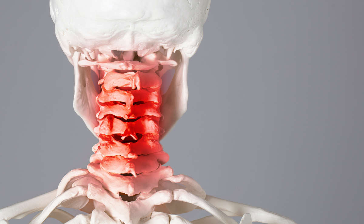
Studies have shown that whiplash injuries are mainly associated with neck pain as a major symptom. It is also reported that “neck pain is present in all patients with WAD, but headache is also a prevalent symptom (88%), especially in patients in whom the C2–3 facet joint is implicated as a cause of pain.
Deans and his team estimated neck pain occurred in “65% of patients within 6 hours, 93% within 24 hours, and 100% within 72 hours after neck injury”. Evidently, there are many variations depending on many factors including the extent of neck injury, mechanism of injury, direction of the insult, force of the collision, and acceleration force.
Studies pointed that patient with WAD, that were examined within 3 days after the trauma, had a significant increase in pro-inflammatory tumor necrosis factor (TNF)-α and interleukin (IL)-6 and of anti-inflammatory IL-10. These normalized in the following 24 hours.
In one study, the incidence of cervical facet joint pathology in WAD, leading to neck pain was 71%.
The distribution pattern of cervical facetogenic pain varies from level to another. Dwyer and team performed intra-articular facet joint injections in 4 volunteers and 1 patient with neck pain to delineate the area of pain from injection. Injection of the C2–3 joint by capsular distension was associated with upper neck pain that extended into the head (often towards the ear, forehead, vertex, or eye). Injections into the C3–4 joint resulted in pain in the neck extending from the suboccipital region to the lower neck without involving the shoulder. Injection into the C4–5 joint caused a more ‘caudal’ pain, in the top of the shoulder and lower part of the neck. Injection of the C5–6 joint resulted in pain radiating into the lower neck, top of the scapula, and shoulder above the level of the scapular spine that was distinguishable from pain extending caudally to the scapular spine from irritation of the C6–7 joint.
To further study cervical facet joint kinematics and injury mechanisms during simulated whiplash injuries, Pearson and his team came up with the idea to emphasize on studying “facet joint compression, facet joint sliding, and capsular ligament strain at all cervical levels during multiple whiplash simulation accelerations”. He concluded that the cervical facet joint components may be at risk for injury due to facet joint compression during a rear-impact accelerations of 3.5 g and above and added that capsular ligaments are also at risk for injury at higher accelerations.
Pearson and his team demonstrated that peak facet joint compression was the greatest at C4–C5, where it reached a maximum of 2.6 mm during the 5 g simulation. He added that increases over physiologic limits were initially observed during the 3.5 g simulation. He also postulated that peak facet joint sliding and capsular ligament strains were largest in the lower cervical spine and increased with impact acceleration. Capsular ligament strain reached a maximum of 39.9% at C6–C7 during the 8 g simulation.